Sai Deep Eye Clinic

Cataract Treatment in Vileparle and Santacruz

Cataract Diagnosis & Treatment

What is Pterygium?

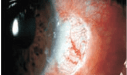
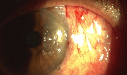
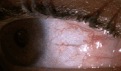
Pterygium is also known as Surfer’s eye. It is an extra growth that develops on the conjunctiva or the mucous membrane that covers the sclera (white part of the eye). It usually grows from the nasal side of the conjunctival.

mass growing on cornea

Pterygium Symptom Checker

What Causes Pterygium?

  • Dryness in the eyes is one of the biggest causes of Pterygium.
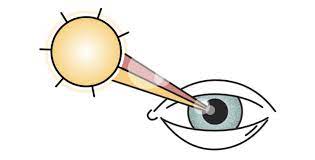
  • Pterygium causes include exposure to ultraviolet rays for a long time.
  • It may be caused due to the dust.
pterygium causes (1)

Diagnosis

  • Slit lamp examination
  • A visual Acuity test- It involves reading letters on an eye chart.
  • Corneal Topography –it is used to measure the curvature changes in your cornea.
  • Photo Documentation- It involves taking pictures to track the growth rate of Pterygium.

Treatment

    • Medical:
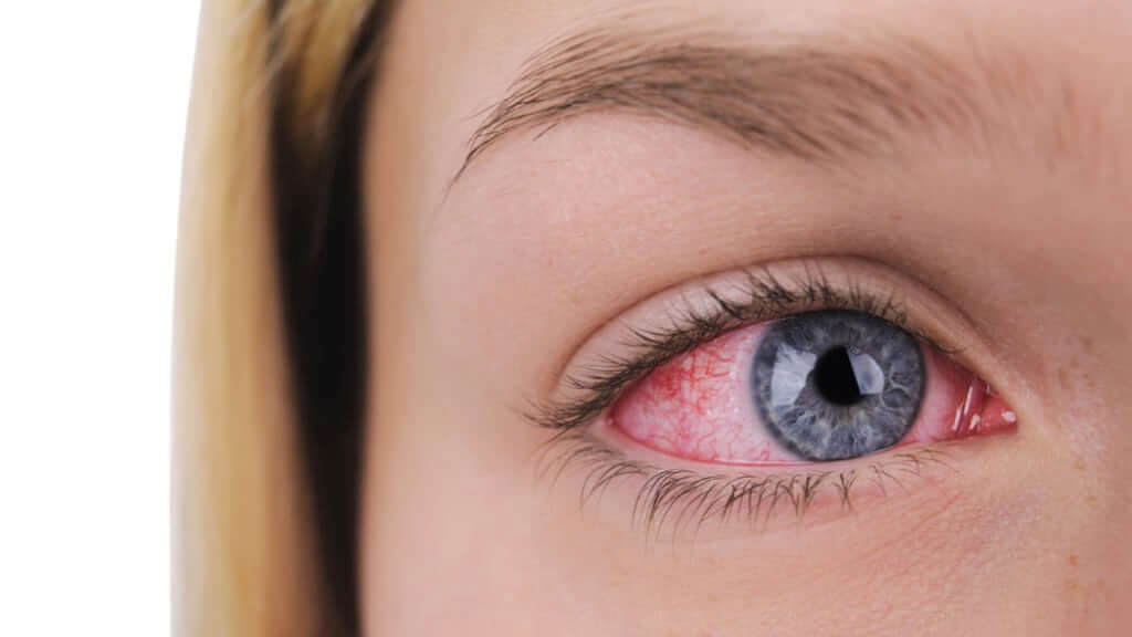
If the Pterygium is leading to symptoms like irritation or redness, the doctor will prescribe eye drops to reduce the inflammation.
    • Surgical:
If the Pterygium symptoms are worsening and the drops is not offering any relief. Your eye doctor will recommend surgery to remove the pterygium.

Treatment

  • Medical:
  • If the Pterygium is leading to symptoms like irritation or redness, the doctor will prescribe eye ointment to reduce the inflammation.

  • Surgical:
  • If the Pterygium symptoms are worsening and the ointment is not offering any relief. Your eye doctor will recommend surgery to remove the pterygium.

Pterygium Reviews

I did my cataract surgery from sai deep
eye clinic for both eyes and it was very successful. I have got clear vision and I am very happy with the services.

Ermelinda P Fernandes

I did my mother’s cataract surgery, the
services are excellent and would truly recommend this clinic for eye related problems…

agnel paul

My mum came here for her cataract
surgery. The staff is very helpful and co operative. Even the staff is very knowledgeable regarding the services they provide. More over the doctors Nitin and Nikhil are very humble and very best at what they do. Will definitely recommend my friends and family

Rinisha Smile

Frequently Asked Questions

What happens during a pterygium surgery?
Pterygium surgery is a relatively low-risk and efficient procedure performed in eye hospitals. The steps involved include:
  • Initially,the patient is given an injection around the eye to numb the eye and prevent discomfort. The surgical area is thoroughly cleaned to minimize infection risks.
  • The surgeon carefully removes the pterygium along with the conjunctival tissue.
  • To prevent future pterygium growth, a membrane tissue graft is placed in the removed area.
What is the ‘Bare Sclera Technique’?

An alternative approach to treating pterygium is the “bare sclera technique”, where the surgeon removes the pterygium without replacing it with new tissue. This technique allows the eye’s white area to heal naturally but carries a higher risk of pterygium regrowth compared to grafting.

What are the complications of pterygium surgery?
Like any surgical procedure, pterygium surgery has potential complications. Patients may experience redness, discomfort, and temporary blurriness during recovery. If issues like vision problems, pterygium regrowth, or vision loss arise, immediate consultation with an ophthalmologist is essential.
What is the difference between in fibrin glue and sutures?

After pterygium removal, surgeons may use either sutures or fibrin glue to secure the conjunctival tissue graft. The key distinctions between these methods are:

  • Sutures are dissolvable but can lead to more post-surgery discomfort and extended healing.
  • Fibrin glue reduces discomfort and shortens recovery time but carries a risk of disease transmission and is costlier.
Surgical


Pterygium involves removal of the abnormal tissue from the sclera and cornea of the eye. Today’s techniques offer a significantly higher success rate than conventional surgery.

1. Pterygium Excision with Bare Sclera:- In traditional “bare sclera” pterygium removal, the underlying white of the eye is left exposed. Healing occurs over two to four weeks with mild to moderate discomfort. Unfortunately, “bare sclera” pterygium surgery has a high rate of re-growth; this occurs in up to 50% of patients. In many cases, the pterygium grows back larger that its original size. Over the years, surgeons have used several different techniques to lessen the likelihood of pterygium recurrence, including radiation treatment and the use of “antimetabolite” chemicals that prevent growth of tissue. Each of these techniques has risks that potentially threaten the health of the eye after surgery, including persistent epithelial defects (ulceration in the surface of the eye), and corneal melting.


2. Pterygium Excision with Conjunctival Autograft with Sutures:- Most corneal specialists today perform pterygium surgery with a conjunctival autograft because of a reduced risk of recurrence.

In this technique, the pterygium is removed, and the cornea regains clarity. However, the gap in the mucous membrane (conjunctiva) tissue, where the pterygium was removed, is filled with a transplant of tissue that has been painlessly removed from underneath the upper eyelid. Although the procedure requires more surgical skill than traditional surgery, this “auto-graft” (self-transplant) helps prevent re-growth of the pterygium by filling the space where abnormal tissue would have re-grown. In
conventional autograft surgery, stitches are used to secure the graft in place on the eye. These can cause discomfort for several weeks. The autograft is held in place with tiny stitches that may dissolve after a few weeks or can be removed in the surgeon’s office. Stitches on the eye frequently cause discomfort, however, after pterygium/autograft surgery.

3. Pterygium Excision with Conjunctival Autograft with Fibrin Glue :- No-stitch surgery is made possible by the use of modern tissue adhesive. Composed of clotting proteins normally found in human blood, tissue adhesive allows the surgeon to secure a conjunctival autograft in seconds rather than minutes. After about one week the tissue adhesive dissolves with no residue, leaving the eye to heal comfortably.

Surgical

Pterygium involves removal of the abnormal tissue from the sclera and cornea of the eye. Today’s techniques offer a significantly higher success rate than conventional surgery.

1. Pterygium Excision with Bare Sclera:-

In traditional “bare sclera” pterygium removal, the underlying white of the eye is left exposed. Healing occurs over two to four weeks with mild to moderate discomfort. Unfortunately, “bare sclera” pterygium surgery has a high rate of re-growth; this occurs in up to 50% of patients. In many cases, the pterygium grows back larger that its original size. Over the years, surgeons have used several different techniques to lessen the likelihood of pterygium recurrence, including radiation treatment and the use of “antimetabolite” chemicals that prevent growth of tissue. Each of these techniques has risks that potentially threaten the health of the eye after surgery, including persistent epithelial defects (ulceration in the surface of the eye), and corneal melting.

2. Pterygium Excision with Conjunctival Autograft with Sutures:-

Most corneal specialists today perform pterygium surgery with a conjunctival autograft because of a reduced risk of recurrence.

In this technique, the pterygium is removed, and the cornea regains clarity. However, the gap in the mucous membrane (conjunctiva) tissue, where the pterygium was removed, is filled with a transplant of tissue that has been painlessly removed from underneath the upper eyelid. Although the procedure requires more surgical skill than traditional surgery, this “auto-graft” (self-transplant) helps prevent re-growth of the pterygium by filling the space where abnormal tissue would have re-grown. In
conventional autograft surgery, stitches are used to secure the graft in place on the eye. These can cause discomfort for several weeks. The autograft is held in place with tiny stitches that may dissolve after a few weeks or can be removed in the surgeon’s office. Stitches on the eye frequently cause discomfort, however, after pterygium/autograft surgery.

3. Pterygium Excision with Conjunctival Autograft with Fibrin Glue :-

No-stitch surgery is made possible by the use of modern tissue adhesive. Composed of clotting proteins normally found in human blood, tissue adhesive allows the surgeon to secure a conjunctival autograft in seconds rather than minutes. After about one week the tissue adhesive dissolves with no residue, leaving the eye to heal comfortably.

Scroll to Top