


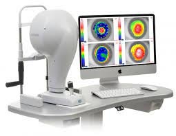


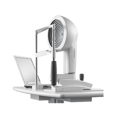
Wavelight Topolyzer Vario
- It is a Placido disc based Corneal Topographer to evaluate the corneal curvature prior to LASIK/Refractive procedures.
- Planning of various refractive procedures and crosslinking procedures for keratoconus is based on this instrument.
- Helps in monitoring the corneal curvature post refractive/crosslinking procedures.
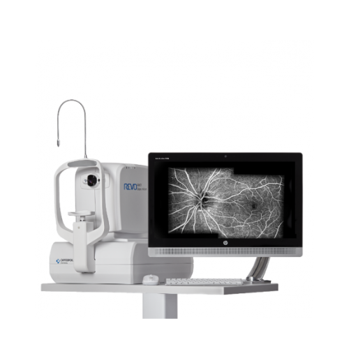
OPTOPOL Revo FC
Optical Coherence Tomography(oct)
- Fully automated and easy to use
- Ultra high speed
- Pachymetry and Epithelial Thickness help in Refractive Surgery Assessment as well in detection of Corneal Ectatic Conditions like Keratoconus
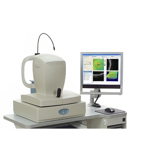
Optovue RTVue
Optical Coherence Tomography
- Gold Standard in Epithelial mapping due to second to none accuracy and precision
- Pachymetry and Epithelial Thickness help in Refractive Surgery Assessment as well in detection of Corneal Ectatic Conditions like Keratoconus
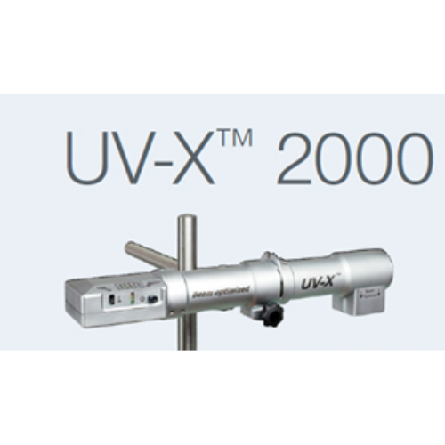
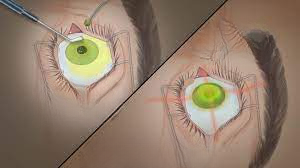
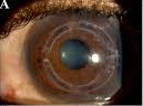
PESCHKE UVX Crosslinking machine
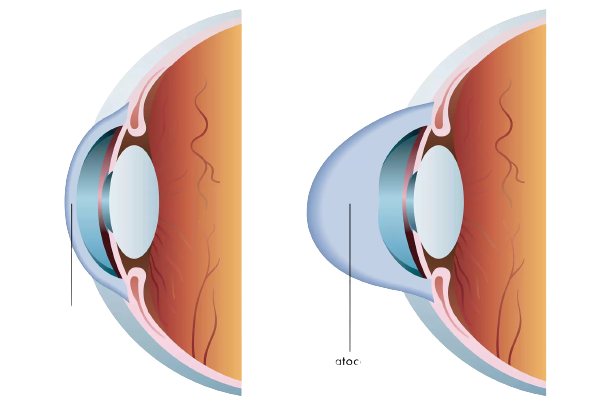
Corneal cross-linking procedure has become the standard procedure for treating patients with progressive keratoconus and other ectatic corneal diseases.
It helps in halting the progression of these conditions by strengthening the weak cornea.
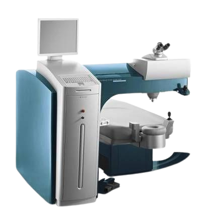
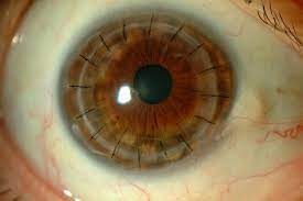
Alcon Wavelight FS 200(Femtosecond) laser
- Ultrafast
- Bladefree treatment
- Painless
- Rapid Recovery
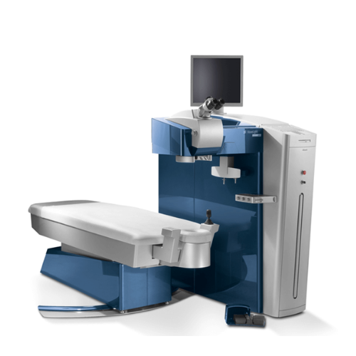
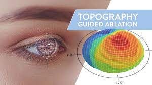
Alcon Wavelight Excimer 500 laser
- Gold standard of excimer laser treatment
- Best in Class Safety
- Fastest Number Correction
- Painless
- Extremely Accurate and Precise
- Predictable Results
OPTOPOL Revo FC
Optical Coherence Tomography(oct)
- Fully automated and easy to use
- Ultra high speed
- OCT is like an ultrasonography of the retina to pick up lesions in different layers of the retina.
- OCT ONH (Glaucoma) helps in precise diagnosis and monitoring of glaucoma progression over time
- Pachymetry and Epithelial Thickness help in Refractive Surgery Assessment as well in detection of Corneal Ectatic Conditions like Keratoconus
Optovue RTVue
Optical Coherence Tomography
- Gold Standard in Epithelial mapping due to second to none accuracy and precision
- OCT is like an ultrasonography of the retina to pick up lesions in different layers of the retina.
- OCT ONH (Glaucoma) helps in precise diagnosis and monitoring of glaucoma progression over time
- Pachymetry and Epithelial Thickness help in Refractive Surgery Assessment as well in detection of Corneal Ectatic Conditions like Keratoconus
Wavelight Topolyzer Vario
- It is a Placido disc based Corneal Topographer to evaluate the corneal curvature prior to LASIK/Refractive procedures.
- Planning of various refractive procedures and crosslinking procedures for keratoconus is based on this instrument.
- Helps in monitoring the corneal curvature post refractive/crosslinking procedures.
PESCHKE UVX Crosslinking machine
Corneal cross-linking procedure has become the standard procedure for treating patients with progressive keratoconus and other ectatic corneal diseases.
It helps in halting the progression of these conditions by strengthening the weak cornea.
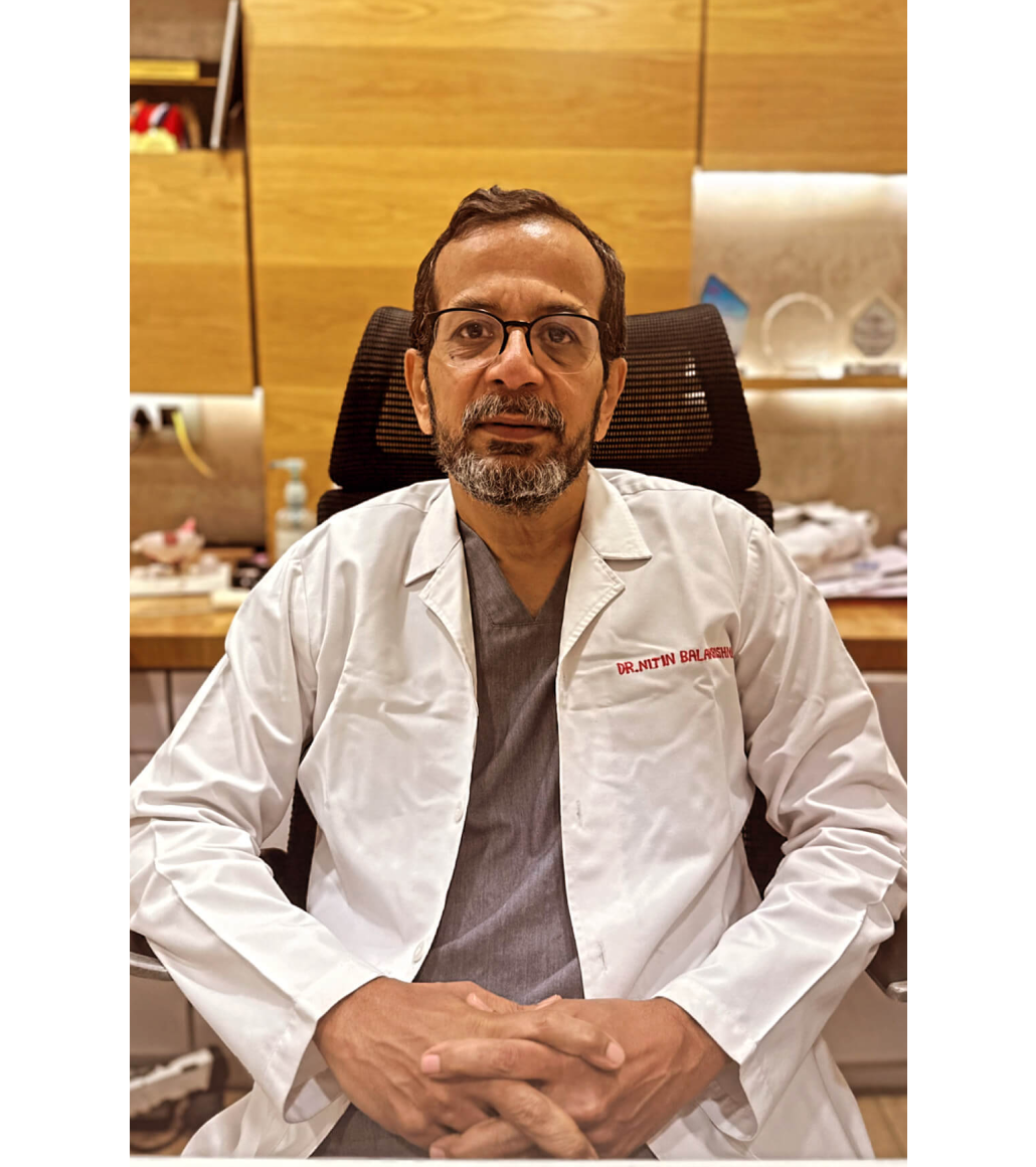
Dr. Nitin
Balakrishnan
Cataract & Refractive Surgeon

Dr. Nikhil Nitin Balakrishnan
Cataract & Refractive Surgeon

Dr. Pavitra Patel Balakrishnan
Cataract & Refractive Surgeon