


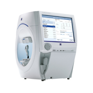
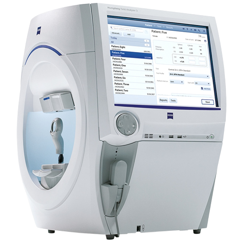
Zeiss Humphrey Field Analyzer 3
- Visual Field Testing for the purposes of screening, monitoring, and assisting in the diagnosis and management of ocular diseases such as glaucoma and related neurological disorders.
- Easy to perform
- Extremely repeatable
- Progression mapping :- Provides information about stability/progression of disease
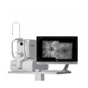
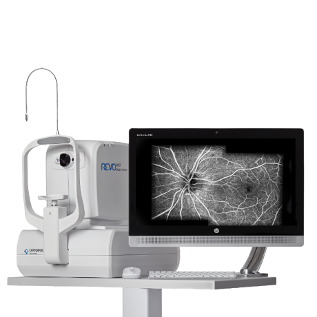
OPTOPOL Revo FC OPTICAL COHERENCE TOMOGRAPHY(OCT)
- OCT ONH (Glaucoma) helps in precise diagnosis and monitoring of glaucoma progression over time
- Pachymetry and Epithelial Thickness help in Refractive Surgery Assessment as well in detection of Corneal Ectatic Conditions like Keratoconus
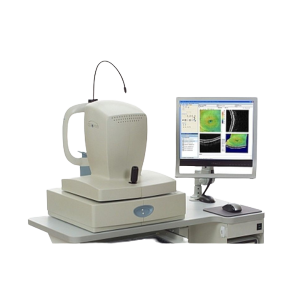
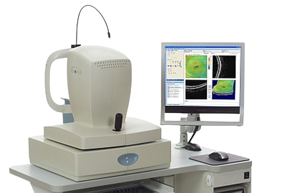
Optovue RTVue OPTICAL COHERENCE TOMOGRAPHY
- Gold Standard in Epithelial mapping due to second to none accuracy and precision
- OCT ONH (Glaucoma) helps in precise diagnosis and monitoring of glaucoma progression over time
- Pachymetry and Epithelial Thickness help in Refractive Surgery Assessment as well in detection of Corneal Ectatic Conditions like Keratoconus
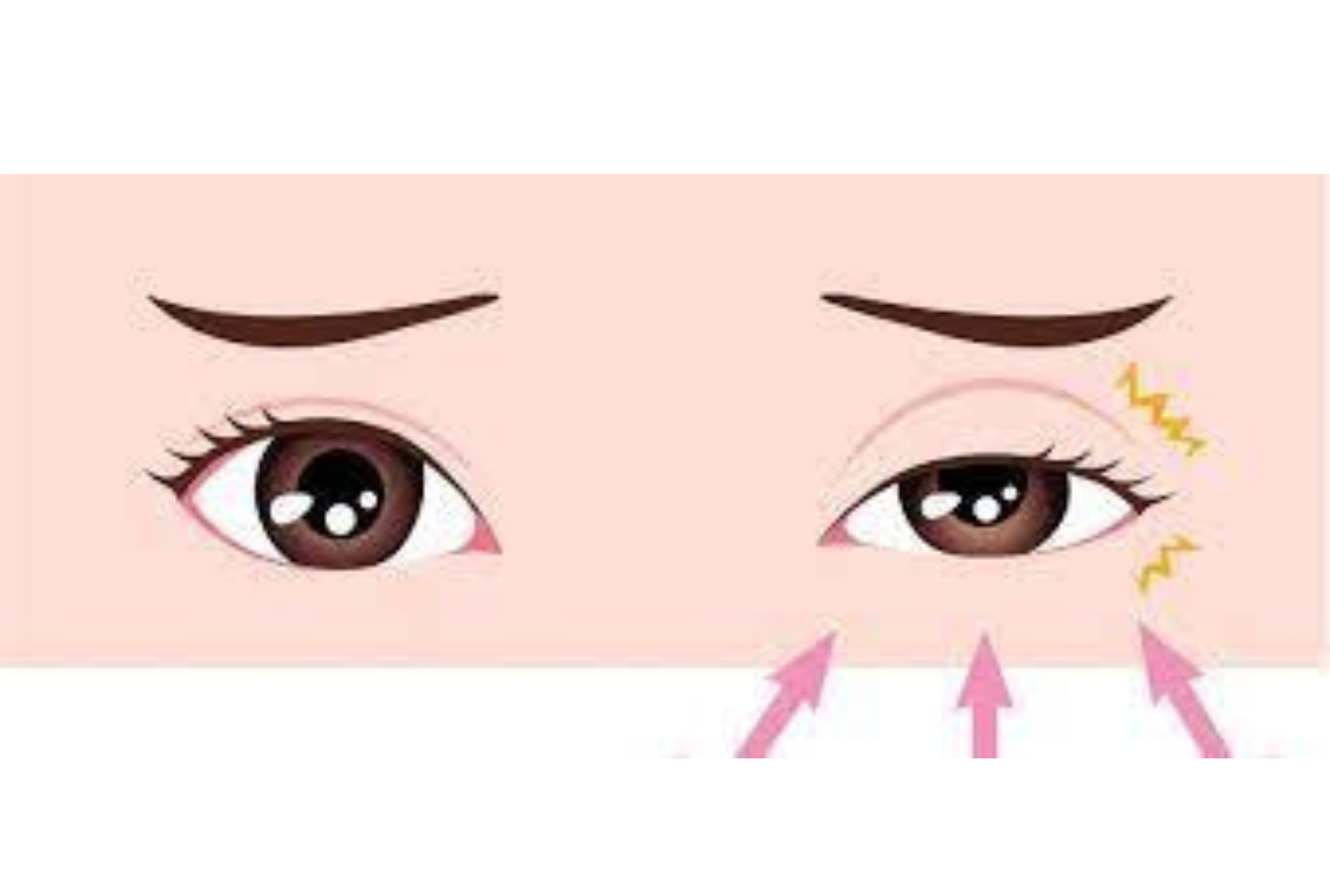
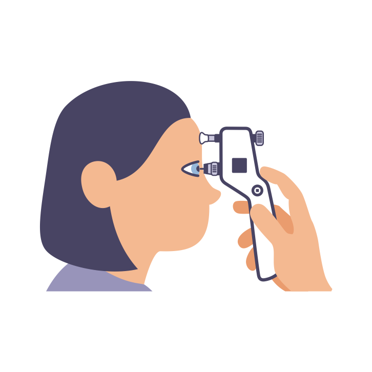 Tonometry is a diagnostic procedure utilized to gauge the intraocular pressure (IOP), which is the fluid pressure inside the eye. This test holds significance as it aids the doctor in assessing the potential risk of glaucoma. For individuals already diagnosed with glaucoma, tonometry is crucial in understanding the risk of disease progression and determining the effectiveness of treatment.
 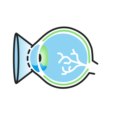 Gonioscopy is a painless examination of the anterior chamber of your eye, which allows the doctor to determine whether the drainage angle, where fluid exits the eye, is open or closed. This test is vital as it enables your doctor to diagnose glaucoma accurately and customize the most suitable treatment plan for your condition.
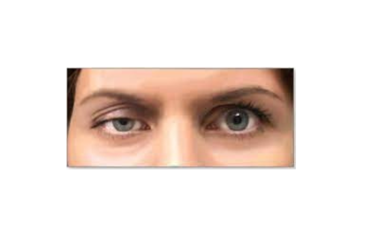
 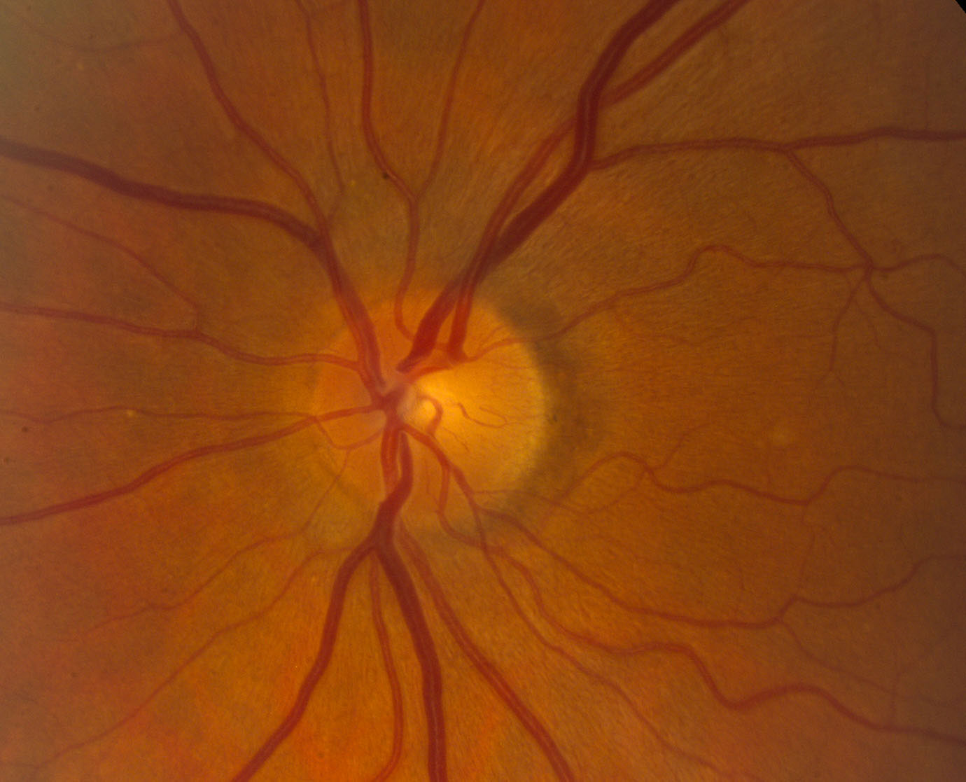 This diagnostic procedure allows the doctor to assess the damage to your optic nerve caused by glaucoma. By using eye drops to dilate the pupil, the doctor can obtain a clear view of the optic nerve, examining its shape and color through your eye.
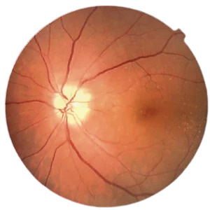
  This diagnostic method assists in assessing the thickness of the cornea, a crucial factor in determining the Corrected IOP (Intraocular Pressure).
 This method aids in the assessment of initial glaucomatous alterations on the Optic Nerve head. It provides information about the thinning of the Retinal Nerve Fiber Layer (RNFL) in different quadrants and changes in the Ganglion Cell Layer (GCC).
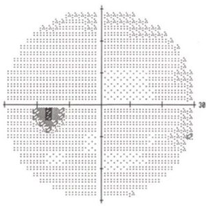
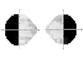
The Perimetry, also known as the Visual Field test, creates a comprehensive map of your entire field of vision. By examining your peripheral or side vision, which is usually impacted first by glaucoma, your doctor can diagnose and track the progression of the condition. The test results are crucial in assessing the severity of glaucoma, the extent of vision loss, as well as any damage to the visual pathways in the brain and other optic nerve-related disorders.


Tonometry is a diagnostic procedure utilized to gauge the intraocular pressure (IOP), which is the fluid pressure inside the eye. This test holds significance as it aids the doctor in assessing the potential risk of glaucoma. For individuals already diagnosed with glaucoma, tonometry is crucial in understanding the risk of disease progression and determining the effectiveness of treatment.
Gonioscopy is a painless examination of the anterior chamber of your eye, which allows the doctor to determine whether the drainage angle, where fluid exits the eye, is open or closed. This test is vital as it enables your doctor to diagnose glaucoma accurately and customize the most suitable treatment plan for your condition.
This diagnostic procedure allows the doctor to assess the damage to your optic nerve caused by glaucoma. By using eye drops to dilate the pupil, the doctor can obtain a clear view of the optic nerve, examining its shape and color through your eye.
This diagnostic method assists in assessing the thickness of the cornea, a crucial factor in determining the Corrected IOP (Intraocular Pressure).
This method aids in the assessment of initial glaucomatous alterations on the Optic Nerve head. It provides information about the thinning of the Retinal Nerve Fiber Layer (RNFL) in different quadrants and changes in the Ganglion Cell Layer (GCC).
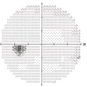
The Perimetry, also known as the Visual Field test, creates a comprehensive map of your entire field of vision. By examining your peripheral or side vision, which is usually impacted first by glaucoma, your doctor can diagnose and track the progression of the condition. The test results are crucial in assessing the severity of glaucoma, the extent of vision loss, as well as any damage to the visual pathways in the brain and other optic nerve-related disorders.
 The Perimetry, also known as the Visual Field test, creates a comprehensive map of your entire field of vision. By examining your peripheral or side vision, which is usually impacted first by glaucoma, your doctor can diagnose and track the progression of the condition. The test results are crucial in assessing the severity of glaucoma, the extent of vision loss, as well as any damage to the visual pathways in the brain and other optic nerve-related disorders.
Dr. Nitin
Balakrishnan
Cataract & Refractive Surgeon

Dr. Nikhil Nitin Balakrishnan