Sai Deep Eye Clinic

Cataract Treatment in Vileparle and Santacruz

Cataract Diagnosis & Treatment

Glaucoma Specialist in Mumbai

What is Glaucoma?

Glaucoma refers to a group of conditions that damages the optic nerve, located at the rear of the eye, responsible for transmitting visual signals to the brain for visualization. This damage can potentially cause blindness. The primary cause of this optic nerve damage in glaucoma is often an abnormally elevated pressure on the nerve, which may eventually lead to vision loss. Glaucoma is recognized as the leading cause of blindness in adults. Some forms of glaucoma may not exhibit noticeable symptoms, making it challenging to detect the condition until it reaches an advanced stage, as its effects progress gradually over time.
What-is-Glaucoma-1024x1024-removebg-preview

What Is The Symptoms Of Glaucoma?


Causes of Glaucoma

  • Build-up of aqueous humor inside the eye
  • Genetic reasons
  • Birth defects
  • Blunt or chemical injury
  • Acute eye infection
  • Blockage by blood vessels inside the eye
  • Inflammatory conditions
  • In rare cases, previous eye surgeries

Types of Glaucoma

PRIMARY OPEN ANGLE GLAUCOMA

The most prevalent form of glaucoma is known as primary open-angle glaucoma. In this type, the drainage passage for the fluid within the eye remains open, allowing fluid outflow. However, there may be a flaw in the drainage pathway, resulting in increased resistance to fluid drainage, leading to heightened eye pressure. In a small percentage of cases, increased fluid production can also contribute to the elevated pressure. This type of glaucoma develops slowly and does not initially present any symptoms. It first affects the side vision, gradually progressing towards the center of vision. Due to this gradual progression, many individuals may remain unaware of their condition until they experience significant vision loss that affects their central vision.

Risk factors for primary open angle glaucoma are:

  • Age more than 40 years
  • Race – African Americans
  • Family history of glaucoma
  • Systemic diseases such as diabetes mellitus, hypertension, heart disease
  • High myopia or those with a high minus power
ANGLE CLOSURE GLAUCOMA OR CLOSED ANGLE GLAUCOMA

Angle closure glaucoma, also known as closed angle glaucoma, is the second most common type of glaucoma and is characterized by narrow drainage channels in the eye. If the angle gradually closes over time, it is referred to as chronic angle closure, while a sudden closure of the angle leads to an acute angle closure attack. Acute angle closure glaucoma presents as a medical emergency. During an acute attack, the patient experiences symptoms such as eye pain, nausea, vomiting, redness, blurred vision, and colored halos due to a rapid increase in eye pressure. Immediate treatment by an eye specialist is essential in such cases.

Risk factors for angle closure glaucoma:

  • Age more than 40 years
  • Race – East Asians
  • Family history of glaucoma
  • Women
  • High hypermetropia or those with a high plus power
NORMAL TENSION GLAUCOMA /LOW TENSION GLAUCOMA

Normal tension glaucoma, also referred to as low tension glaucoma, is a type of glaucoma where damage to the optic nerve can occur despite the eye pressure being within the normal range. The exact cause of this condition is not fully understood, but it is believed to stem from either an exceptionally delicate optic nerve that can be harmed even with normal eye pressure or reduced blood flow to the optic nerve. This type of glaucoma is often asymptomatic, and individuals typically do not experience any visual complaints until the disease reaches a very advanced stage.

Risk factors for normal tension glaucoma are:

  • Family history of normal tension glaucoma
  • Race – Japanese ancestry
  • Thinner corneas
  • Heart disease
  • Migraine and peripheral vascular disease
CONGENITAL GLAUCOMA (CHILDHOOD GLAUCOMA)

Glaucoma during infancy and childhood is a relatively uncommon but serious set of diseases that can jeopardize vision. Infants and young children with glaucoma are usually brought for an eye examination because either their parents or pediatricians have observed some peculiarities in their eyes. These signs may include whitish discoloration and enlargement of the eyes, excessive tearing, a tendency to keep the eyes closed, and sensitivity to light. Glaucoma in children may arise if there’s a family history of glaucoma, if the parents have had a consanguineous (close blood relation) marriage, or due to abnormal development during pregnancy.

This type of glaucoma occurs due to an irregular flow of intraocular fluid within the eye caused by a blockage or dysfunction of the trabecular meshwork, which are the drainage canals in the eye with a mesh-like structure. Alternatively, in certain instances, the abnormal drainage system can be a consequence of another eye or body condition, leading to what is known as secondary glaucoma.

Congenital glaucoma is identified through a comprehensive eye examination, typically conducted under sedation in the operating theater for infants and children below the age of 3. Early detection and treatment of congenital glaucoma can help slow down the progression of the condition. Microsurgery is often used to address structural defects in straightforward cases of congenital glaucoma. For more complex cases, a combination of medication and surgery is employed for treatment.

SECONDARY GLAUCOMA

Secondary glaucoma refers to specific types of glaucoma in which the underlying reason for increased eye pressure leading to optic nerve damage and vision loss can be identified. Various factors contribute to secondary glaucoma, such as the prolonged and unregulated use of steroids, severe diabetic retinopathy, eye injuries, inflammation of the eye (uveitis), or advanced stages of cataract.

If you suspect having any of these risk factors, it is important to undergo an eye examination. Remember to inform your eye doctor about your specific risk factors, which will assist them in determining the frequency of your eye check-ups. The type of treatment prescribed will be based on whether you have open-angle or angle-closure glaucoma.

PIGMENTARY GLAUCOMA
Pigmentary Glaucoma is classified as a type of secondary open-angle glaucoma. It occurs when pigment granules from the back of the iris, the colored part of the eye, enter the clear fluid produced inside the eye. Over time, these tiny pigment granules flow towards the drainage canals of the eye, gradually obstructing them and leading to increased eye pressure. As more pigments are released, patients may experience blurred vision during physical exertion or exercise. This type of glaucoma is frequently observed in young males who have high nearsightedness and wear strong minus power lenses.
TRAUMATIC GLAUCOMA

Secondary open-angle glaucoma can be a result of eye injury. Traumatic glaucoma can manifest immediately after the injury or emerge years later. It can be triggered by blunt trauma, which bruises the eye, or by penetrating injuries. In most cases of blunt trauma, the elevated eye pressure is temporary. However, there are instances where the drainage canals in the eye sustain damage and permanent scarring occurs. This scarring obstructs fluid flow and may lead to glaucoma even years after the initial injury.

Furthermore, conditions like myopia (nearsightedness), previous eye injury, infections, or past surgeries can increase the susceptibility of the eye to severe injuries.

PSEUDOEXFOLIATIVE GLAUCOMA

Pseudoexfoliative Glaucoma, a type of secondary open-angle glaucoma, occurs when a flaky material similar to dandruff peels off from the outer layer of the eye’s lens. This material accumulates in the angle between the cornea and iris, potentially blocking the eye’s drainage system and leading to increased eye pressure. Pseudoexfoliative Glaucoma is frequently observed in individuals of Scandinavian descent.

Individuals diagnosed with exfoliative glaucoma commonly experience more instances of elevated eye pressure, greater fluctuations in pressure, and higher peak pressures compared to patients with other forms of glaucoma. Managing this type of glaucoma with medical treatment alone is typically more challenging. Patients with exfoliative glaucoma necessitate aggressive medical therapy and are more likely to require laser or surgical interventions. Regular and frequent visits to their eye doctor are often essential to closely monitor the progression of the disease.

NEOVASCULAR GLAUCOMA

A type of secondary open-angle glaucoma can be triggered by the atypical growth of new blood vessels on the iris and across the eye’s drainage channels.

Neovascular glaucoma is invariably linked to other conditions, most commonly diabetes and hypertension. It never develops in isolation. The emergence of new blood vessels obstructs the normal drainage of eye fluid through the trabecular meshwork, resulting in elevated eye pressure. This type of glaucoma advances swiftly and poses significant challenges in terms of treatment.

Glaucoma Risk Factors

You are most likely to get glaucoma if you:

  • Are you over 60 years of age
  • Have high internal eye pressure
  • Have a family member diagnosed with glaucoma
  • Have certain conditions like diabetes, heart conditions, sickle cell anaemia and high blood pressure.
  • Have thin corneas
  • Have extreme conditions of nearsightedness or farsightedness
  • Have had eye injuries, surgeries
  • Taking corticosteroid medications for a long time

Glaucoma Technology

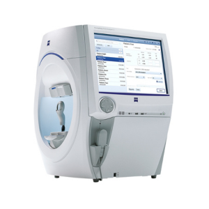
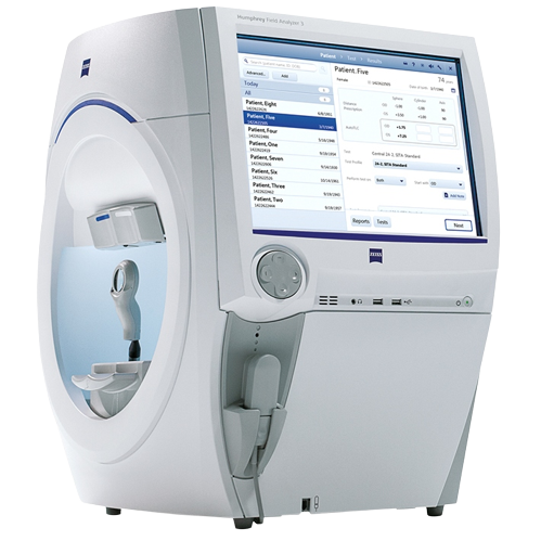
Zeiss Humphrey Field Analyzer 3

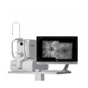
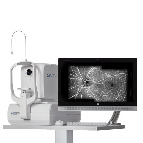
OPTOPOL Revo FC OPTICAL COHERENCE TOMOGRAPHY(OCT)

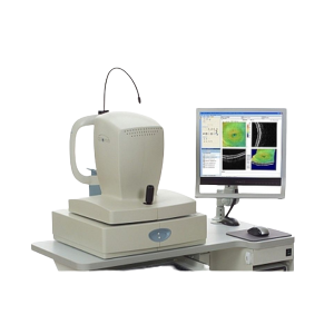
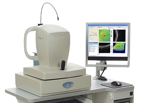
Optovue RTVue OPTICAL COHERENCE TOMOGRAPHY

Zeiss_Humphrey_Field_Analyzer_3-removebg-preview
OPTOPOL_Revo_FC-removebg-preview
Optovue_RTVue-removebg-preview
  • Visual Field Testing for the purposes of screening, monitoring, and assisting in the diagnosis and management of ocular diseases such as glaucoma and related neurological disorders.
  • Easy to perform
  • Extremely repeatable
  • Progression mapping :- Provides information about stability/progression of disease
  • OCT ONH (Glaucoma) helps in precise diagnosis and monitoring of glaucoma progression over time
  • Pachymetry and Epithelial Thickness help in Refractive Surgery Assessment as well in detection of Corneal Ectatic Conditions like Keratoconus
  • Gold Standard in Epithelial mapping due to second to none accuracy and precision
  • OCT ONH (Glaucoma) helps in precise diagnosis and monitoring of glaucoma progression over time
  • Pachymetry and Epithelial Thickness help in Refractive Surgery Assessment as well in detection of Corneal Ectatic Conditions like Keratoconus

Glaucoma Diagnosis

Regular eye examinations are crucial for maintaining eye health. At the age of 40, it is recommended to undergo a baseline eye screening to detect any early signs of eye disease or vision changes that may begin to emerge. Based on the results of this screening, your eye doctor will advise you on the frequency of follow-up exams to ensure optimal eye care.

If you possess risk factors for glaucoma, such as diabetes, hypertension, or a family history of the condition, it is advisable to schedule an appointment with an eye doctor promptly. They can assess your situation and recommend the appropriate frequency for your eye examinations.

A thorough glaucoma assessment involves a series of tests that aid in detecting the existence of glaucoma and monitoring its progression during subsequent follow-ups.

Once a thorough evaluation of the patients is completed, doctors engage in discussions regarding the most suitable treatment options available. These options may include topical eye drops, oral medications, or interventional procedures such as lasers or surgeries. Patients are encouraged to inquire and express any concerns they may have, and doctors make an effort to address all their queries with care and attentiveness.

The utilization of these novel investigative techniques enables us to identify the disease and its progression at an earlier stage, leading to improved patient management.

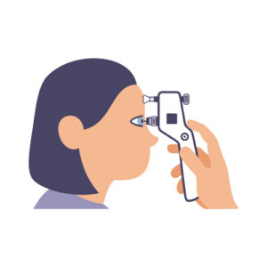
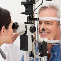
Tonometry is a diagnostic procedure utilized to gauge the intraocular pressure (IOP), which is the fluid pressure inside the eye. This test holds significance as it aids the doctor in assessing the potential risk of glaucoma. For individuals already diagnosed with glaucoma, tonometry is crucial in understanding the risk of disease progression and determining the effectiveness of treatment.

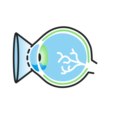
Gonioscopy is a painless examination of the anterior chamber of your eye, which allows the doctor to determine whether the drainage angle, where fluid exits the eye, is open or closed. This test is vital as it enables your doctor to diagnose glaucoma accurately and customize the most suitable treatment plan for your condition.
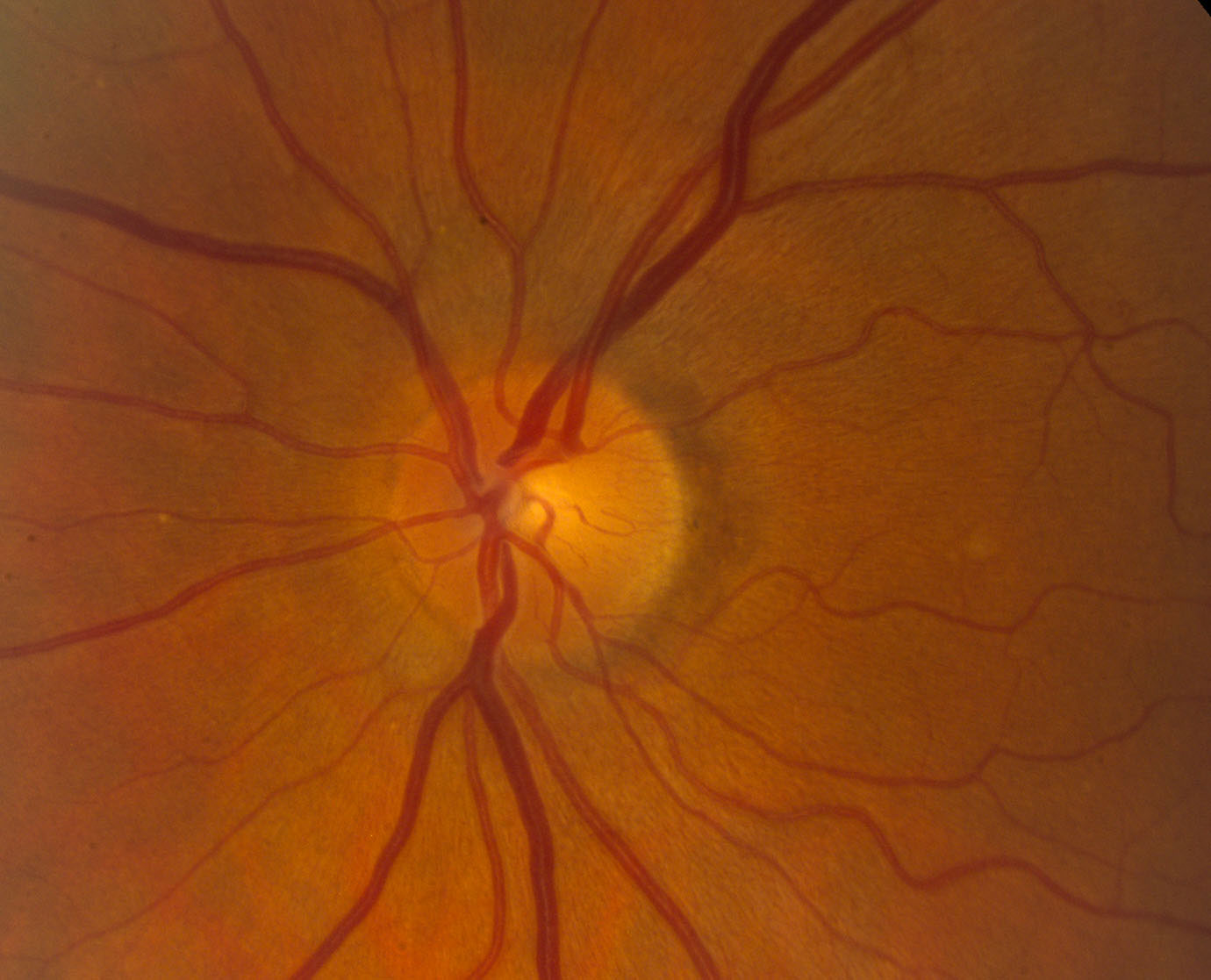
This diagnostic procedure allows the doctor to assess the damage to your optic nerve caused by glaucoma. By using eye drops to dilate the pupil, the doctor can obtain a clear view of the optic nerve, examining its shape and color through your eye.
This diagnostic method assists in assessing the thickness of the cornea, a crucial factor in determining the Corrected IOP (Intraocular Pressure).
This method aids in the assessment of initial glaucomatous alterations on the Optic Nerve head. It provides information about the thinning of the Retinal Nerve Fiber Layer (RNFL) in different quadrants and changes in the Ganglion Cell Layer (GCC).

Visual Field test
The Perimetry, also known as the Visual Field test, creates a comprehensive map of your entire field of vision. By examining your peripheral or side vision, which is usually impacted first by glaucoma, your doctor can diagnose and track the progression of the condition. The test results are crucial in assessing the severity of glaucoma, the extent of vision loss, as well as any damage to the visual pathways in the brain and other optic nerve-related disorders.

TREATMENT FACILITIES AT SAI DEEP EYE CLINIC

aao-C50
The damage caused by Glaucoma is irreversible, but early treatment and regular examinations can effectively slow down or prevent vision loss, particularly in the early stages. The treatment for Glaucoma primarily revolves around reducing the Intraocular Pressure (IOP) in your eyes. Depending on the severity of the condition, treatment options may include eye drops, oral medications, laser procedures, surgery, or a combination of these approaches.

GLAUCOMA TREATMENT OPTIONS

Medical Treatment

  • Your physician will initiate the treatment by recommending specific eye drops to lower your eye pressures. These drops work either by enhancing fluid drainage from the eye or by reducing the production of fluid in the eye. Depending on the target eye pressure, you may be prescribed more than one type of eye drop.

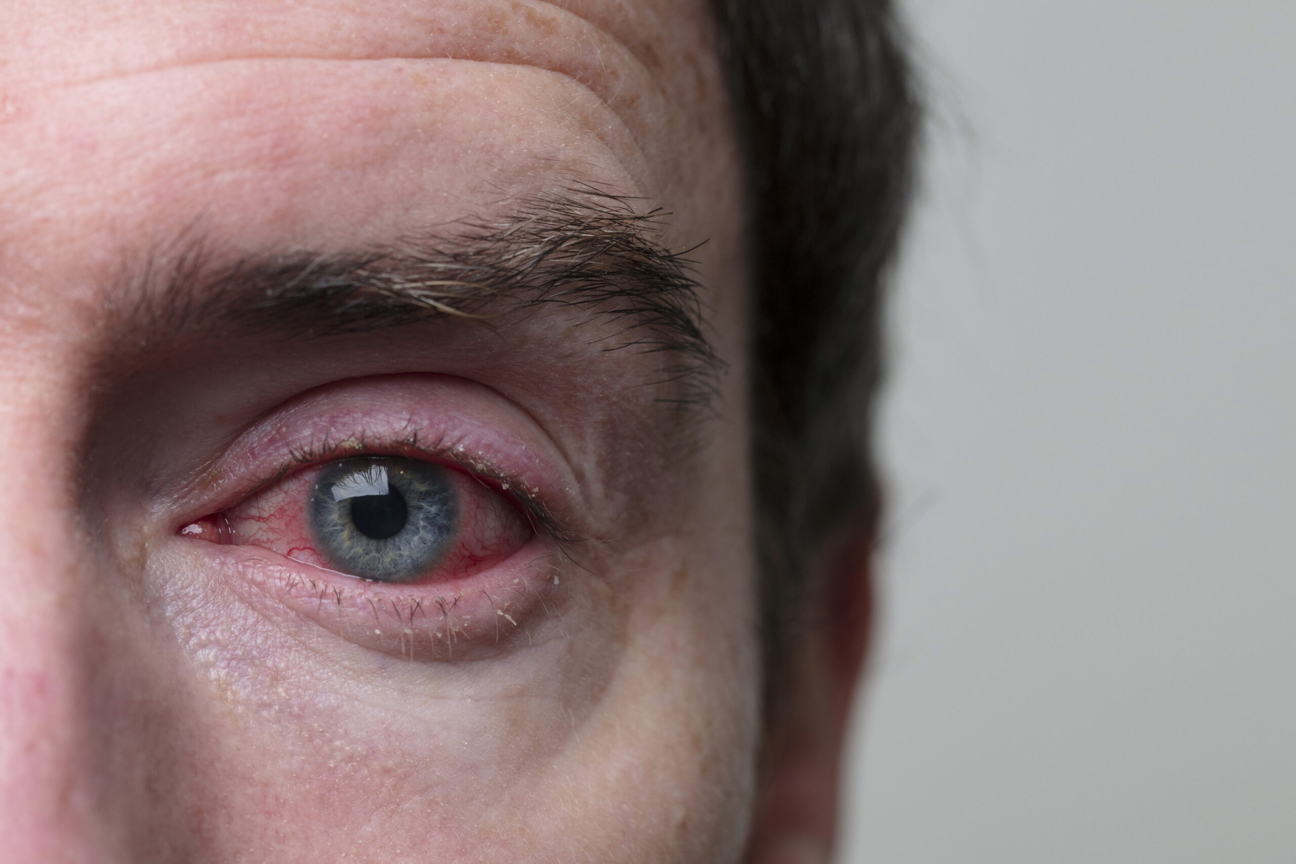
  • Certain eye drops may lead to mild eye irritation, a burning sensation, redness, or changes in eye pigmentation. In rare cases, these eye drops can also cause systemic side effects when absorbed into the body. If you experience discomfort or concerns regarding your eye drops, it is important to discuss them with your doctor.

Laser Peripheral Iridotomy (LPI)

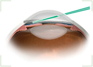
Laser peripheral iridotomy is the conventional treatment for closed-angle glaucoma and eyes susceptible to this condition. During this procedure, a laser beam is used to create an aperture in the iris, providing an alternative pathway for fluid drainage from the eye.
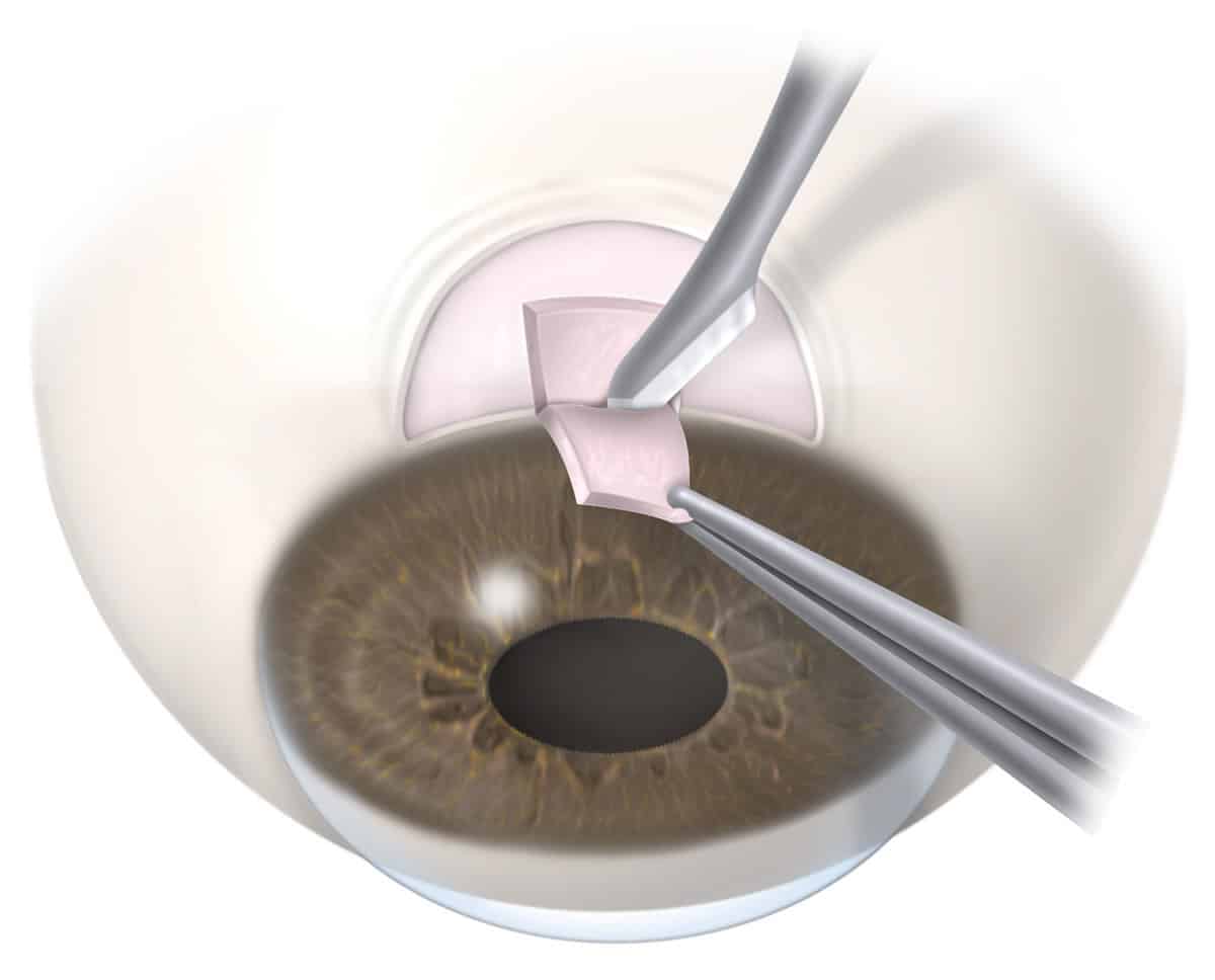
Trabeculectomy

During this surgical procedure, a small opening is created in the eye's wall, protected by a flap that functions like a trap door. Controlled drainage of fluid from the inside of the eye occurs through this opening, resulting in the formation of a small "bleb" beneath the upper eyelid. This surgical intervention is employed to reduce eye pressure when medical treatment or laser therapy has not been successful.

Glaucoma Drainage Device (GDD)

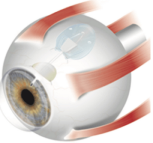
If glaucoma medications have proven ineffective in managing intraocular pressure, your doctor may propose tube shunt surgery. This procedure involves redirecting the aqueous humor (fluid inside the eye) from the eye to an external reservoir.

The Ahmed Glaucoma Valve (AGV) and the Aurolab Aqueous Drainage Device (AADI) are frequently utilized glaucoma drainage devices crafted from silicon polypropylene. Resembling a miniature computer mouse, these devices consist of a tube that enters the eye, while the remaining part of the implant rests on the eye's surface, beneath the conjunctiva and concealed by the eyelid. The majority of tube shunt procedures are effective and can prevent glaucoma from progressing to blindness.

Transscleral Cyclophotocoagulation (TSCPC) - Traditional and Micropulse Cyclo G6 Laser
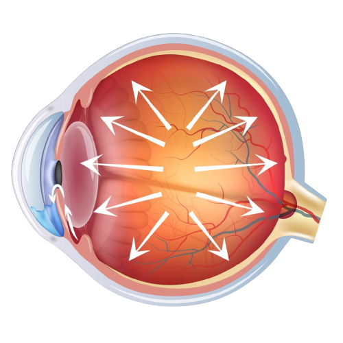
A diode laser, which does not require any incisions, is employed to target the ciliary body, responsible for producing the clear fluid inside the eye. By causing its destruction, the laser procedure effectively reduces the eye pressure.

This procedure is commonly used for managing uncontrollable increases in eye pressure. Conventional Transscleral Cyclophotocoagulation (TSCPC) is generally reserved for cases with poor or limited visual potential, whereas micropulse TSCPC can be utilized in eyes with good visual potential. Your doctor may suggest this treatment if you have experienced multiple unsuccessful glaucoma surgeries or to alleviate pain in ablind eye.

This procedure is conducted as a day care process in the operation theatre under local anesthesia. A probe is positioned on the eye's surface, and laser energy is administered at various points. TSCPC is generally an effective and relatively safe procedure.

Glaucoma Prevention

In the modern era, individuals are well-informed about how lifestyle choices impact their general health, including glaucoma. Lifestyle choices can have some influence on eye pressure and the risk of glaucoma development, but there is a limited amount of proven research on this topic among glaucoma patients.
8358930

Exercise

Studies suggest that maintaining a regular exercise routine and leading an active lifestyle may lower the risk of developing glaucoma. Engaging in activities that increase your heart rate by about 20 to 25 beats per minute, such as brisk walking, can help reduce intraocular pressure (IOP). While aerobic exercises have been observed to cause a temporary decrease in IOP, their effect on glaucoma patients has not been thoroughly examined. Additionally, swimming is believed to enhance the optic nerve’s resilience to an increase in intraocular pressure.
Diet

Diet

Consuming a diet abundant in green leafy vegetables is believed to reduce intraocular pressure.

Foods such as shellfish, tuna, nuts, and seeds that are high in DHA (docosahexaenoic acid) are beneficial for maintaining a healthy retina.

Incorporating spinach, broccoli, and sprouts into your diet, which contain lutein and zeaxanthin, can also help prevent oxidative damage to the optic nerve.

What to Avoid?

Exercise avoid

Exercises to be Avoided

Lifting heavy weights can lead to a temporary rise in intraocular pressure, so individuals with glaucoma are advised to avoid lifting excessive weights. Additionally, certain yoga asanas, especially those involving a head-down position like sheershasana, have been found to elevate eye pressure. Hence, caution is recommended for glaucoma patients when performing such poses.
food to be avoid

Foods to be Avoided

Foods that are high in trans-fats, like deep-fried items, hinder the proper functioning of omega-3 fatty acids and can lead to an elevation in eye pressure.
young-man-disclaim-cigarette-beer-toned-photo-cigarettes-alcohol-60719048-e4fXX2vKg-transformed

Habit Forming Substances

Caffeine, alcohol, and tobacco have adverse effects on eye pressure. Excessive consumption of caffeine has been shown to elevate the risk of glaucoma. Regular intake of alcohol and smoking cigarettes can also be harmful to the optic nerve.
concentrated-musician-playing-his-instrument_150588-101

Musical Instruments to be Avoided

Playing the trumpet and saxophone have been linked to a rise in eye pressure.

Best Glaucoma Specialist in Mumbai

drkartik

Dr. Kartik
Panikkar

Glaucoma Surgeon

Glaucoma Reviews

Lorem ipsum dolor sit amet, consectetur adipiscing elit. Integer nec purus at urna eleifend consectetur. Vestibulum ante ipsum primis in faucibus orci luctus et ultrices posuere cubilia Curae; Sed dapibus metus id felis sollicitudin, ac elementum quam convallis.

Karan Kothari

Lorem ipsum dolor sit amet, consectetur adipiscing elit. Integer nec purus at urna eleifend consectetur. Vestibulum ante ipsum primis in faucibus orci luctus et ultrices posuere cubilia Curae; Sed dapibus metus id felis sollicitudin, ac elementum quam convallis.

Tan

Lorem ipsum dolor sit amet, consectetur adipiscing elit. Integer nec purus at urna eleifend consectetur. Vestibulum ante ipsum primis in faucibus orci luctus et ultrices posuere cubilia Curae; Sed dapibus metus id felis sollicitudin, ac elementum quam convallis.

Sakshi Shetty

Frequently Asked Questions

WHAT IS CONSIDERED AS NORMAL EYE PRESSURE?
There is no specific standard for normal eye pressure as it can vary based on multiple factors. On average, your eye pressure should fall within the range of 10-21mmHg, but it is also influenced by various elements such as gender, age, ethnicity, family history, and corneal thickness, among others.
SHOULD I GET A GLAUCOMA EVALUATION DONE?
Every person over the age of 40 should undergo an annual eye examination. However, glaucoma screening becomes essential if there is a family history of glaucoma, presence of systemic diseases like diabetes or hypertension, high myopia, a history of eye trauma, or prolonged use of steroids.
HOW FREQUENTLY SHOULD I COME FOR A GLAUCOMA FOLLOW UP?
Your doctor will determine the frequency of follow-up appointments, considering the severity of the condition. It is crucial to adhere to these regular follow-ups to prevent the progression of the disease.
IS GLAUCOMA A CURABLE DISEASE?
Unfortunately, the current nerve damage cannot be reversed. Nevertheless, timely intervention and consistent follow-up appointments can help prevent the disease from advancing further.
CAN I DRIVE IF I HAVE GLAUCOMA?
The majority of individuals with glaucoma can continue to drive. Your eligibility to drive will be determined by the extent of your visual field loss. Your doctor will conduct regular visual field tests to assess this aspect.
WHAT SHOULD I DO IF I HAVE MISSED A FOLLOW UP?
It is essential to adhere to the regular and timely administration of medications until you can promptly visit your doctor.
WHAT IS THE CORRECT TECHNIQUE FOR INSTILLING GLAUCOMA EYE DROPS?
In order to reduce systemic absorption, close your eyes for one to two minutes after applying the eye drops. Afterwards, gently press the corner of your eye near your nose for one to two minutes to close the tear duct. Wipe away any excess eye drops from your eyelid. If you are using more than one eye drop, ensure a 5-minute interval between each drop.
CAN I HAVE SYSTEMIC SIDE EFFECTS WITH GLAUCOMA MEDICATIONS?
It is uncommon, but in some cases, systemic absorption of the medication may lead to various side effects. These can include dizziness, reduced heart rate, worsening of asthma, impotence, tingling sensations in the extremities, fatigue, metallic taste, increased frequency of urination, dry mouth, and others, depending on the specific medication being used. If you encounter any new systemic effects after starting any medication, it is essential to promptly consult your doctor.
WHAT IS THE ANGLE OF THE EYE AND CLOSED ANGLE GLAUCOMA?

The angle refers to the gap between the cornea (clear part) and the iris (colored part) of the eye, which allows the aqueous fluid to drain out of the eye.

Closed angle glaucoma occurs when the angle between the cornea and iris becomes narrow or closes, resulting in a rapid or gradual increase in eye pressure. This elevated pressure can lead to optic nerve damage and potential vision loss.

WHAT SHOULD I EXPECT DURING THE PERIPHERAL IRODOTOMY PROCEDURE?
It is done as an OPD procedure.
  • Before starting the treatment, eye drops are administered to constrict the pupil, a process that may require approximately 1-2 hours.
  • Right before the procedure, numbing drops are applied to the surface of the eye.
  • Afterward, a lens is positioned on the eye to conduct the laser procedure.
  • The procedure typically lasts around 10-15 minutes for both eyes to be completed.
WHAT CAN I EXPECT AFTER THE PERIPHERAL IRODOTOMY PROCEDURE?
Temporary blurring of vision, redness in the eye, or heightened sensitivity to light may occur, but these effects typically resolve within a day. Following the procedure, anti-inflammatory drops are prescribed, and the eye pressure is evaluated after one week.
WILL PERIPHERAL IRODOTOMY IMPROVE MY VISION?
Not in every case. During an acute angle closure attack, patients may encounter blurred vision caused by fluid accumulation in the cornea, the clear part of the eye. Peripheral iridotomy helps alleviate the blockage of fluid outflow and clears the cornea, thus restoring vision to its pre-attack state. However, the main purpose of the procedure is to preserve the existing vision and prevent glaucoma progression.
WHAT ARE THE RISKS OF PERIPHERAL IRODOTOMY?
In the vast majority of cases, the procedure has minimal to no risks. The documented risks include the closure of iridotomy, which may necessitate re-treatment, excessive bleeding (especially in patients taking blood thinners), cataract progression, and, in rare instances, retinal and choroidal detachments. Your doctor will thoroughly discuss your individual risk profile with you and take appropriate precautions as needed.
WHAT ARE THE RISKS OF TRABECULECTOMY PROCEDURE?
While rare, there is a possibility of severe complications arising from the surgery, including infection, intraocular bleeding, vision loss, and eye pressure that is too low or too high. As a result, regular follow-up appointments to monitor the eye pressure, surgical wound, bleb (if applicable), and disease progression are crucial even after the surgery.
WILL I HAVE TO CONTINUE MY GLAUCOMA EYEDROPS POST TRABECULECTOMY SURGERY?
The majority of patients do not need medications to manage eye pressure after trabeculectomy. However, in some cases, despite the surgical intervention, there may still be progression of the condition. If your doctor determines that the eye pressure is not at an optimal level or if there is ongoing progression, you may be advised to continue using the eye drops.
IS TUBE SHUNT SURGERY SAFE?

The procedure is generally safe and effective for controlling eye pressure. However, there are potential complications, such as infection, tube extrusion, corneal decompensation, and implant failure, which can occur even years after the surgery. Therefore, caution and regular follow-up appointments are essential.

It is crucial to adhere to regular follow-up appointments as recommended by your doctor to achieve the best possible outcomes.

WHAT ARE THE RISKS ASSOCIATED WITH TSCPC PROCEDURE?
The procedure is generally safe, but in rare cases, there might be complications such as inflammatory reactions, pain, redness in the eye, conjunctival scarring, extremely low eye pressure, and further vision loss.
Dr. Kartik Panikkar

Glaucoma Surgeon

MBBS, DNB -Ophthalmology, Fellowship in Glaucoma ,FICO ,FAICO,FRCS

Scroll to Top