


Cataract Treatment in Vileparle and Santacruz
Cataract Diagnosis & Treatment
Macular Hole Surgery
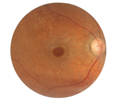
What is a Macular Hole?
As a whole, it can enlarge over time at the distorted area of the eye- portion causing serious side effects. This full-thickness disorder requires immediate surgery at its early stage. Although this is a rare vitreoretinal interface disorder; however, it needs surgery right away if the macular hole is getting bigger.
Causes of Macular Hole
We have outlined below a variety of causes that can lead to macular holes.
- Ageing: Ageing is one of the key factors to contribute to the development of a macular hole. As we age, the vitreous gel becomes more liquid and thus becomes prone to shrinkage.
- Eye damage: Another factor contributing to the developemnt of macular hole, are the physical injuries that can occur to the eye due to some accidents or other reason. Even very small injuries can have a major impact on the fragile tissues of the eye.
- Myopia (nearsightedness): People with high degrees of myopia are more likely to develop macular holes. This is because myopia can cause the eyeball to elongate, putting additional strain on the macula.
- Pre-existing Eye Conditions: Conditions like retinal detachment, retinoschisis, and macular pucker can also increase the likelihood of developing macular holes. These conditions can create structural vulnerabilities in the eye, making it more susceptible to damage.
Macular Hole Symptoms
The symptoms of a macular hole can vary from person to person, but they generally include:
- Blurred or Distorted centre Vision: Blurred or distorted centre vision is one of the most common symptoms of a macular hole. Usually here we can observe wavy or curved lines in the field of vision.
- Reduced Colour Perception: Colours may appear less bright due to this condition.
- Central Vision Dark patch: A macular hole can cause a dark patch in your vision. It’s as if you’re staring through a little hole or tunnel, and the quality of your eyesight suffers as a result.
- Difficulty with Reading and Close-Up jobs: Macular holes make it difficult to recognize faces, read far off signs etc.
Who is at Risk of Developing a Macular Hole?
Types of Macular Holes
Diagnosis
The steps of macular hole diagnosis are:
- Visual acuity test: A visual acuity test determines how clear your central vision is. Patients are asked to read letters or symbols at various distances to determine the quality of their central vision.
- Dilated Eye Exam: The doctor uses eye drops to dilate the patient’s pupils, allowing the retina and macula to be examined more thoroughly.
- Optical Coherence Tomography (OCT): This non-invasive imaging technique creates high-resolution cross-sectional images of the macula. It enables a precise examination of the aspects of the macular hole.
Treatment
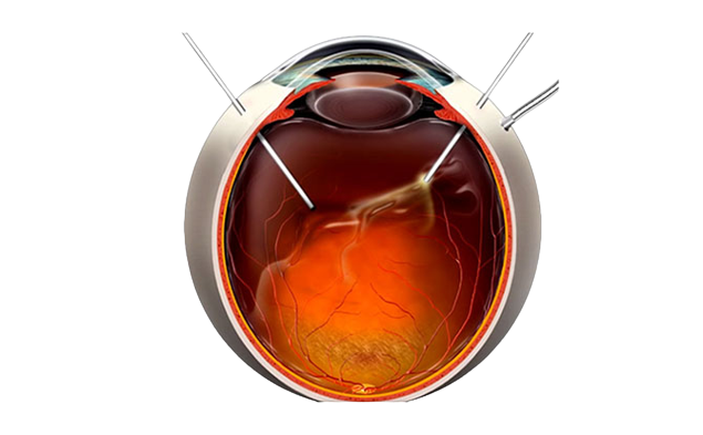
- Vitrectomy: Vitrectomy is a surgical technique in which the vitreous gel in the eye is removed. A gas bubble is then injected to help in the closure of the macular hole. The body absorbs the gas bubble over time, allowing the hole to heal.
- Face-down Posing: Following a vitrectomy, patients may be required to lie face down for an extended period. This position keeps the gas bubble in contact with the macular hole, which can help in healing.
- Recovery and Follow-Up: It is advisable to visit for regular eye checkups and follow-up appointments to ensure complete healing.
While macular holes are a serious problem, they are often curable if appropriate and timely actions are taken. It is important to understand the symptoms and risks to understand early issues.
Frequently Asked Questions
After the surgery, the patient has the option to decide between lying down or sitting in one position with the support of a headrest. This step is crucial as it ensures a proper gas sealing effect on the macular hole.
The surgeon creates an incision in the eye to carefully introduce medical instruments for removing the fluid. Furthermore, they initiate the removal of small tissues or membranes near the macular hole using forceps. This action prevents the macular hole from closing during the procedure, ensuring the smooth progression of the surgery.
During the final phase of macular hole treatment, a sterile gas is substituted for the fluid within the eye to maintain a specific pressure on the macular hole until it heals adequately.
Normally, you will be prescribed medications like Tylenol or similar pain relievers. However, if these medications do not provide relief, it is important to inform your doctor. Also, mild or even intense redness is a common occurrence, but it will gradually diminish over time.
To prevent any issues, it is advisable to avoid high altitudes or elevations that might cause the gas bubble to expand beyond its normal size. This expansion could potentially lead to eye damage. Therefore, it is better to refrain from flying until the gas bubble is completely absorbed.
In many cases, lamellar holes can be associated with other medical conditions such as Vitreomacular traction, Epi-retina membrane, Cystoid macular edema, and others. To ensure appropriate and timely treatment, your ophthalmologist will examine your eyes for all these mentioned conditions.