

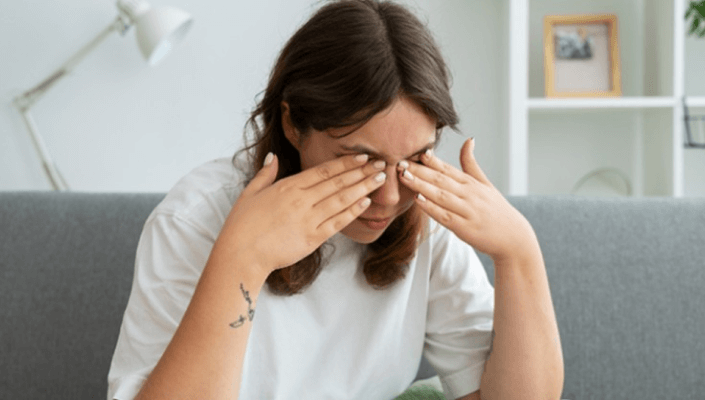


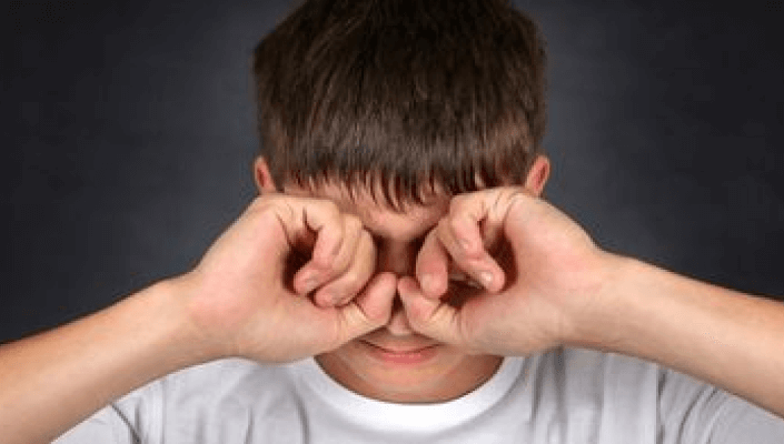
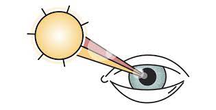

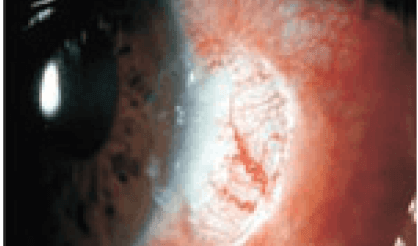
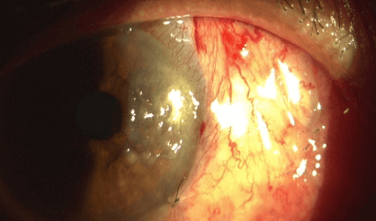
Pterygium involves removal of the abnormal tissue from the sclera and cornea of the eye. Today’s techniques offer a significantly higher success rate than conventional surgery.
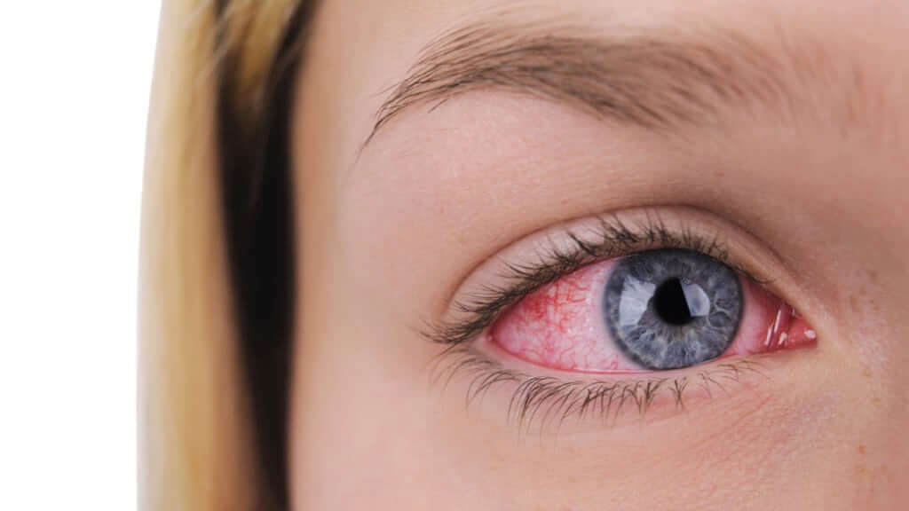
1. Pterygium Excision with Bare Sclera:- In traditional “bare sclera” pterygium removal, the underlying white of the eye is left exposed. Healing occurs over two to four weeks with mild to moderate discomfort. Unfortunately, “bare sclera” pterygium surgery has a high rate of re-growth; this occurs in up to 50% of patients. In many cases, the pterygium grows back larger that its original size. Over the years, surgeons have used several different techniques to lessen the likelihood of pterygium recurrence, including radiation treatment and the use of “antimetabolite” chemicals that prevent growth of tissue. Each of these techniques has risks that potentially threaten the health of the eye after surgery, including persistent epithelial defects (ulceration in the surface of the eye), and corneal melting.
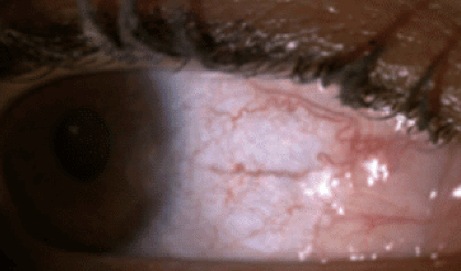
2. Pterygium Excision with Conjunctival Autograft with Sutures:- Most corneal specialists today perform pterygium surgery with a conjunctival autograft because of a reduced risk of recurrence.
In this technique, the pterygium is removed, and the cornea regains clarity. However, the gap in the mucous membrane (conjunctiva) tissue, where the pterygium was removed, is filled with a transplant of tissue that has been painlessly removed from underneath the upper eyelid. Although the procedure requires more surgical skill than traditional surgery, this “auto-graft” (self-transplant) helps prevent re-growth of the pterygium by filling the space where abnormal tissue would have re-grown. In
conventional autograft surgery, stitches are used to secure the graft in place on the eye. These can cause discomfort for several weeks. The autograft is held in place with tiny stitches that may dissolve after a few weeks or can be removed in the surgeon’s office. Stitches on the eye frequently cause discomfort, however, after pterygium/autograft surgery.
3. Pterygium Excision with Conjunctival Autograft with Fibrin Glue :- No-stitch surgery is made possible by the use of modern tissue adhesive. Composed of clotting proteins normally found in human blood, tissue adhesive allows the surgeon to secure a conjunctival autograft in seconds rather than minutes. After about one week the tissue adhesive dissolves with no residue, leaving the eye to heal comfortably.
Pterygium involves removal of the abnormal tissue from the sclera and cornea of the eye. Today’s techniques offer a significantly higher success rate than conventional surgery.
1. Pterygium Excision with Bare Sclera:-
In traditional “bare sclera” pterygium removal, the underlying white of the eye is left exposed. Healing occurs over two to four weeks with mild to moderate discomfort. Unfortunately, “bare sclera” pterygium surgery has a high rate of re-growth; this occurs in up to 50% of patients. In many cases, the pterygium grows back larger that its original size. Over the years, surgeons have used several different techniques to lessen the likelihood of pterygium recurrence, including radiation treatment and the use of “antimetabolite” chemicals that prevent growth of tissue. Each of these techniques has risks that potentially threaten the health of the eye after surgery, including persistent epithelial defects (ulceration in the surface of the eye), and corneal melting.
2. Pterygium Excision with Conjunctival Autograft with Sutures:-
Most corneal specialists today perform pterygium surgery with a conjunctival autograft because of a reduced risk of recurrence.
In this technique, the pterygium is removed, and the cornea regains clarity. However, the gap in the mucous membrane (conjunctiva) tissue, where the pterygium was removed, is filled with a transplant of tissue that has been painlessly removed from underneath the upper eyelid. Although the procedure requires more surgical skill than traditional surgery, this “auto-graft” (self-transplant) helps prevent re-growth of the pterygium by filling the space where abnormal tissue would have re-grown. In
conventional autograft surgery, stitches are used to secure the graft in place on the eye. These can cause discomfort for several weeks. The autograft is held in place with tiny stitches that may dissolve after a few weeks or can be removed in the surgeon’s office. Stitches on the eye frequently cause discomfort, however, after pterygium/autograft surgery.
3. Pterygium Excision with Conjunctival Autograft with Fibrin Glue :-
No-stitch surgery is made possible by the use of modern tissue adhesive. Composed of clotting proteins normally found in human blood, tissue adhesive allows the surgeon to secure a conjunctival autograft in seconds rather than minutes. After about one week the tissue adhesive dissolves with no residue, leaving the eye to heal comfortably.