Sai Deep Eye Clinic

Cataract Treatment in Vileparle and Santacruz

Cataract Diagnosis & Treatment

Retinal Surgery

What is Retinal Surgery?

Retinal surgeries are specialized ophthalmic procedures aimed at addressing various retinal disorders and conditions affecting the delicate tissue at the back of the eye. Conditions such as retinal detachment, macular degeneration, diabetic retinopathy, and epiretinal membranes can impair vision and potentially lead to blindness if left untreated. However, thanks to significant advancements in surgical techniques and technology, retinal surgeries have become increasingly successful, offering hope to patients with these sight-threatening conditions.

Retina

Retinal Surgeries

Retinal Surgery

Vitrectomy

Vitrectomy is a sophisticated surgical procedure performed on the eye’s vitreous gel, a transparent, jelly-like substance that fills the space..
Retinal Surgery

Retinal Detachment Repair

Retinal detachment occurs when the retina separates from the underlying tissue, resulting in vision loss..
Macular hole surgery

Macular Hole
Repair

A macular hole is a small break in the macula, the central part of the retina responsible for sharp, detailed vision. Surgery to..
Retinal Surgery

Epiretinal Membrane (ERM) Surgery

An ERM is a thin, fibrous layer that forms on the surface of the macula, leading to distorted vision. Surgical..
Vitrectomy

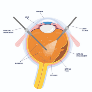
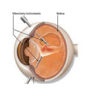
Vitrectomy is a sophisticated surgical procedure performed on the eye’s vitreous gel, a transparent, jelly-like substance that fills the space between the lens and the retina. This delicate surgery is commonly used to treat a variety of sight-threatening conditions affecting the retina and vitreous, such as retinal detachment, macular hole, epiretinal membrane, diabetic retinopathy, vitreous hemorrhage, and more. With the aid of advanced surgical techniques and cutting-edge equipment, vitrectomy has become a crucial intervention in preserving and restoring vision for countless patients worldwide.

Steps of Vitrectomy

  1. Anesthesia: Vitrectomy is typically performed under local anesthesia, with the patient awake but comfortable. In some cases, general anesthesia may be used, especially when the patient requires additional surgical procedures or if they are
    unable to cooperate during the surgery.
  2. Creating Incisions: The surgeon starts by making small, micro-incisions in the sclera, the white outer layer of the eye. These incisions serve as entry points for the surgical instruments used during the procedure.
  3. Removal of Vitreous: Using specialized microsurgical instruments, the surgeon carefully removes the vitreous gel from the eye. The vitreous may be replaced with a gas bubble or silicone oil, depending on the nature of the eye condition being
    treated.
  4. Addressing the Retinal Condition: Once the vitreous is removed, the surgeon can directly access the retina and address the specific retinal condition. This may involve repairing retinal tears or detachment, removing epiretinal membranes, treating diabetic retinopathy, or closing macular holes.
  5. Laser Photocoagulation: During the surgery, the surgeon may use laser photocoagulation to seal retinal tears, cauterize abnormal blood vessels, or treat diabetic retinopathy. Laser technology helps prevent future complications and stabilize the retina.
  6. Gas or Oil Tamponade (Optional): If a gas bubble or silicone oil is used to replace the vitreous, it helps reattach the retina by pushing it against the back of the eye. The gas bubble gradually gets absorbed by the body, while silicone oil may need to be removed in a subsequent procedure once the eye has healed.
  7. Closing Incisions: After completing the necessary interventions, the surgeon closes the micro-incisions using sutures or, in some cases, allows them to self-seal without the need for sutures.
  8. Postoperative Care and Recovery:
    Following vitrectomy, patients are typically monitored for a short period before being discharged. Depending on the specific condition and individual healing progress, the recovery period may vary. Patients are advised to avoid strenuous activities and heavy lifting during the healing process. Regular follow-up visits with the ophthalmologist are crucial to monitor progress, manage any potential complications, and optimize visual outcomes.

Retinal Detachment Repair

Retinal detachment occurs when the retina separates from the underlying tissue, resulting in vision loss. There are several techniques used to repair retinal detachment, such as scleral buckling, pneumatic retinopexy, and vitrectomy. Scleral buckling involves the placement of a silicone band around the eye to push the sclera inward, supporting the reattachment of the detached retina. Pneumatic retinopexy involves injecting a gas bubble into the eye to press the detached retina against the back of the eye, allowing it to reattach naturally.

Macular Hole Repair

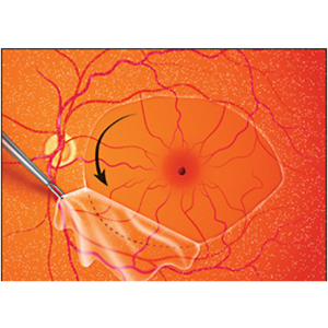
A macular hole is a small break in the macula, the central part of the retina responsible for sharp, detailed vision. Surgery to repair a macular hole typically involves vitrectomy, peeling of the internal limiting membrane, and filling the hole with a gas bubble to promote healing.

Epiretinal Membrane (ERM) Surgery

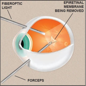
An ERM is a thin, fibrous layer that forms on the surface of the macula, leading to distorted vision. Surgical treatment involves vitrectomy with membrane peeling to remove the ERM and improve vision.

Scroll to Top