

Retinal Vein Occlusion Treatment
Retinal Vein Occlusion?
This eye disorder blocks the flow of the blood by the blood clot in the retina that has no cure if leads to severe complications. Yet you can consult Sai Deep Clinic immediately for RVO treatments to minimize the risk of vision loss. An eye care specialist can prevent the extend of any further complications
There are mainly two types of Retinal vein occlusion:
- Central retinal vein occlusion (CRVO): CRVO develops when the major central vein in the retina, known as the central retinal vein, gets blocked or partially blocked. This blockage might obstruct blood flow from the entire retina.
- Branch retinal vein occlusion (BRVO): BRVO is a condition in which one of the branch veins draining blood from the retina becomes blocked. BRVO, unlike CRVO, affects a smaller, more localised portion of the retina.

Types of Retinal Vein Occulsion - CRVO & BRVO
Central Retinal Vein Occlusion (CRVO)
What central retinal vein occlusion?
The central retinal vein, that is, the primary channel responsible for draining blood from the entire retina, is blocked or partially blocked in CRVO. CRVO can vary in level of severity and impact, and it is commonly divided into two types:
- Non-ischemic CRVO: This consists of some blockage in the central retinal vein but does not result in any significant retinal injury. As a result, mild vision loss is possible, but it is usually not as severe as in ischemic CRVO.
- Ischemic CRVO: The blockage of the veins here is more severe, resulting in a significant reduction in blood flow and oxygen supply to the retina. Ischemic CRVO is linked to an increased risk of visual loss and consequences.
Central retinal vein occlusion symptoms
- Sudden, painless vision loss.
- The appearance of floaters in the visual field.
- In some cases, mild eye pain or discomfort may be experienced.
Central retinal vein occlusion treatment:
Treatment for CRVO frequently involves addressing underlying problems such as high blood pressure or diabetes that may have contributed to the blockage. Complications including macular oedema (swelling of the centre section of the retina) must also be managed. Among the treatment options available are:
- Medication: Anti-VEGF medicines or steroids may be used to treat macular oedema.
- Laser Therapy: Laser therapy may be used to treat aberrant blood vessels in the eye. Surgical intervention may be required in some circumstances.
- Early detection and intervention are crucial in the management of CRVO and the prevention of additional visual loss.
Branch Retinal Vein Occlusion (BRVO):
What is branch retinal vein occlusion?
BRVO is a condition in which one of the branch veins that drain blood from a part of the retina becomes blocked rather than the central vein. Unlike CRVO, BRVO affects only a smaller portion of the retina. BRVO is classified into two subtypes:
- Major BRVO: When one of the main branch veins becomes blocked, it might cause more severe vision problems in the affected area of the retina. Depending on the location and intensity of the blockage, the degree of visual loss may vary.
- Macular BRVO: The macula, the core region of the retina responsible for sharp, central vision, is affected by this type of BRVO. Because of its impact on this vital organ, macular BRVO might cause more pronounced vision problems.
Symptoms :
- Sudden, painless vision loss in the affected area.
- Distortion or visual disturbances within the blocked region.
- Presence of floaters in the visual field.
Branch retinal vein occlusion treatment :
– Medication: Anti-VEGF medicines or steroids may be used to treat macular oedema.
– Laser Therapy: Laser therapy can also be used to treat the unwanted blood vessels in the affected area.
– Regular monitoring is required to ensure that the condition does not deteriorate over time.
As with CRVO, early detection and therapy are critical in controlling BRVO and preserving or regaining eyesight. Individuals who are at risk or having symptoms should see an eye expert right away for a full evaluation and personalised treatment plan. Managing underlying health issues such as hypertension and diabetes is critical for lowering the incidence of retinal vein occlusions.
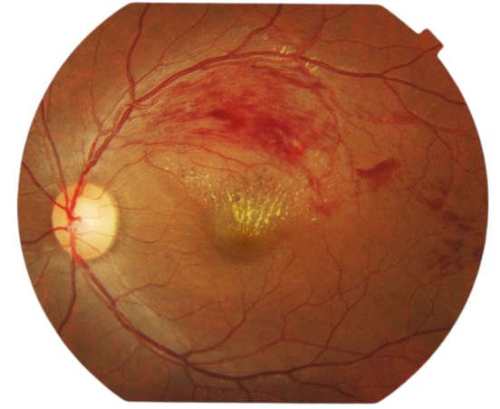
Stages of Branch Retinal Vein Occlusion (BRVO)
Treatment Options for BRVO
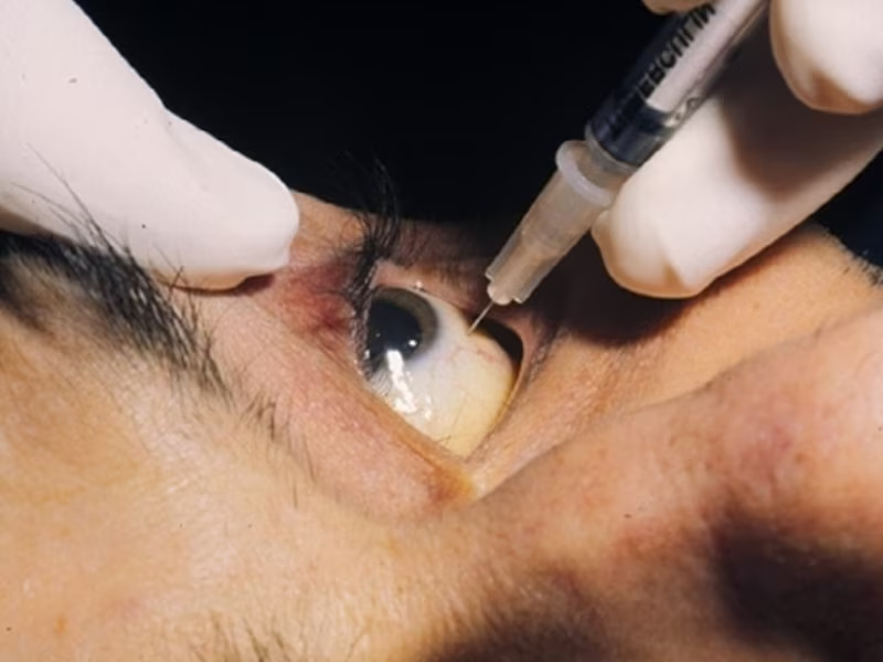
Administering VEGF inhibitors like Lucentis (approved by the FDA), Eylea (also FDA approved), and Avastin (off-label use) through intravitreal injections, or using steroids like triamcinolone and Ozurdex (FDA approved), can aid in sustaining or enhancing vision.
Laser treatment is occasionally employed to address macular edema, but it is currently regarded as a secondary option for most cases, considering visual outcomes and restrictions. For example, if there is substantial retinal hemorrhage, laser treatment cannot be administered until the hemorrhage resolves on its own, which typically takes a few months.
If neovascularization is detected during a follow-up visit, laser treatment can be utilized to shrink these abnormal and delicate vessels, thus reducing the risk of vitreous hemorrhage and vision loss. However, laser treatment cannot guarantee complete prevention of future hemorrhages, but it does substantially lower this risk. In some instances where there is a severe or persistent (non-clearing) vitreous hemorrhage, vitrectomy surgery may be necessary to remove the accumulated blood.